 ◆
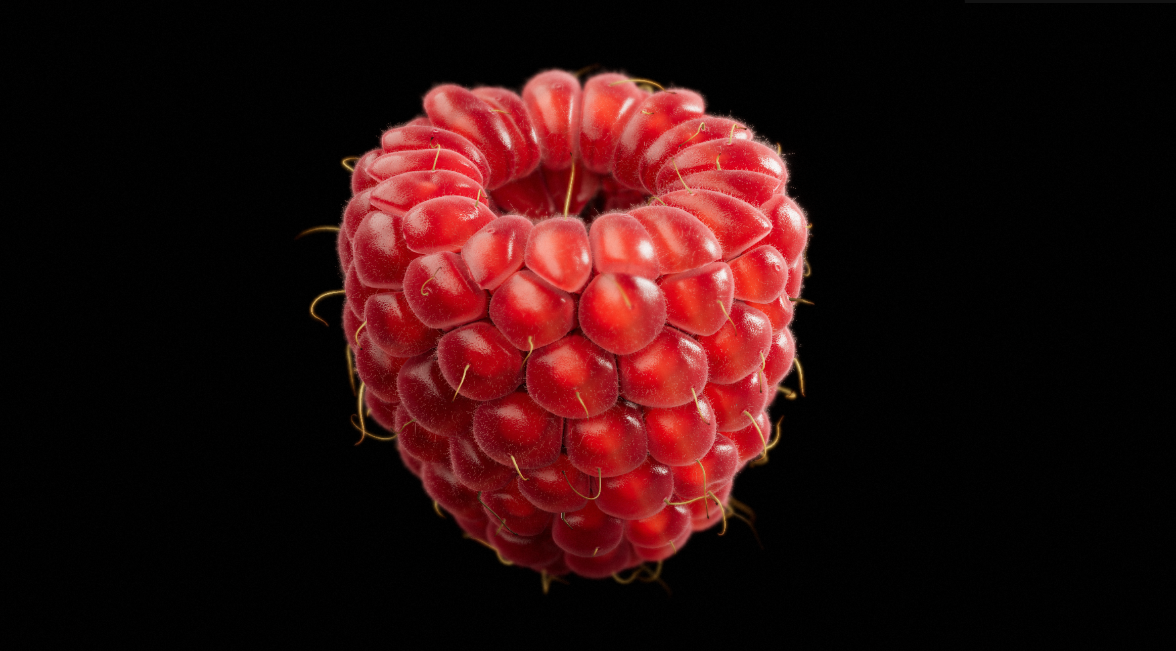
◆Raspberry · Photorealistic 3D
A photorealistic raspberry against a black background, with detailed drupelet surfaces, fine hairs and lighting.
Turn unseen structures into relationships you can explore.
Life-science visualizations connect form, structure and process. Cell models offer a spatial overview. The bacteriophage animation separates recognition, attachment, DNA transfer, synthesis, assembly and release into selectable stages to make their sequence easier to follow.
Teaching models may enlarge membranes, DNA or protein particles and reduce the number of objects. Colors and movement support an explanation rather than reproduce a microscopy image. First distinguish the inside from the outside of a cell, then follow one structure across stages.
◆A photorealistic raspberry against a black background, with detailed drupelet surfaces, fine hairs and lighting.
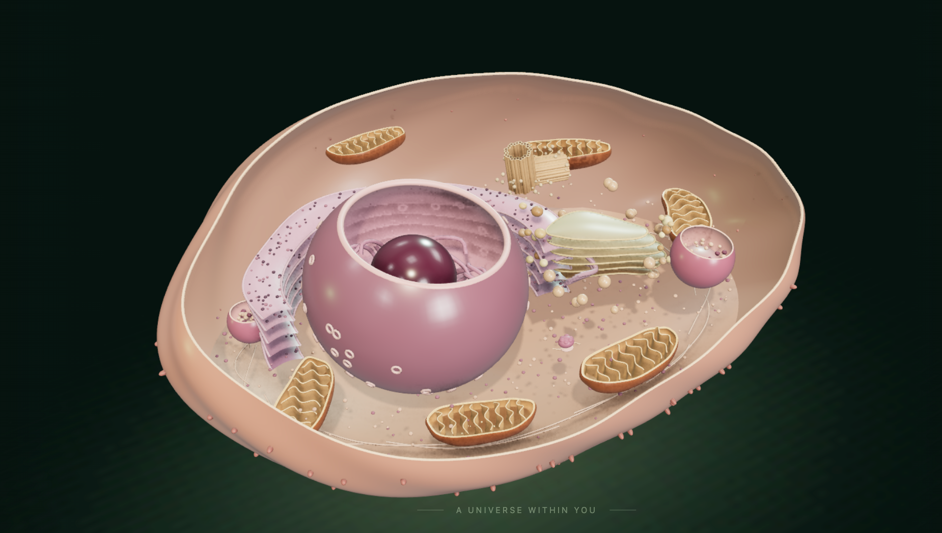 ◆
◆An interactive 3D atlas of plant and human cells: rotate, cut away and inspect organelles, with Chinese and English support.
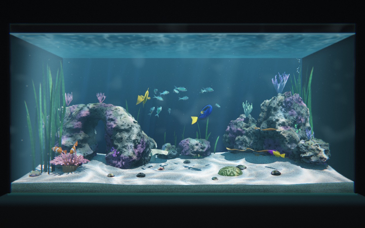 ◆
◆Explore a reef aquarium with tropical fish, underwater lighting and swimming animations. Drag to orbit and scroll to zoom.
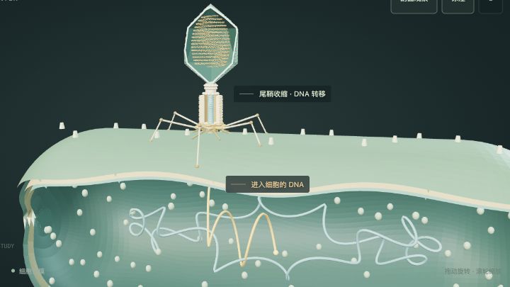 ⬡
⬡Explore T4 infection of E. coli in six interactive steps, from receptor recognition to lysis. Rotate the cutaway to follow DNA into the cell.